Stem cells collected in late pregnancy herald advances in prenatal medicine
A new pioneering approach, developed by researchers at UCL and Great Ormond Street Hospital means human development can be observed in late pregnancy for the first time, raising the possibility of monitoring and treating congenital conditions before birth.
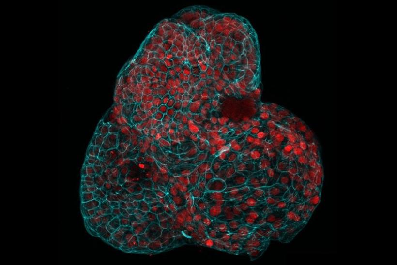
The study, published in Nature Medicine, documents the first time that complex cell models, called organoids, have been grown from human stem cells during an active pregnancy. These ‘mini-organs’ also retain the baby’s biological information.
Organoids provide researchers with invaluable tools to study how organs function both when they are healthy and when impacted by disease.
The researchers say the stem cell organoids will facilitate monitoring of fetal development in late pregnancy, modelling of disease progression and testing of new treatments for diseases such as congenital diaphragmatic hernia (CDH).
In this study, researchers extracted and characterised live cells from amniotic fluid samples taken from 12 pregnancies as part of routine diagnostic testing. They then used single-cell RNA sequencing to identify which tissues these stem cells came from. Stem cells from the lungs, kidneys and intestine were successfully extracted, which were used to grow organoids that had functional features of these tissue types. Because the child would not be touched during the collection process, sampling restrictions would be overcome and the cells would carry the same biological information as the child.
To assess how the organoids might be used in the management of congenital diseases, the team worked with researchers at KU Leuven in Belgium to study the development of babies with CDH, a condition where a hole in the diaphragm means organs like the intestine and liver get displaced into the chest, putting pressure on the lungs and hindering healthy growth.
Organoids from babies with CDH both pre- and post-treatment were compared to organoids from healthy babies to study the biological characteristics of each group. As expected, there were significant developmental differences between the healthy and pre-treatment CDH organoids. But the organoids in the post-treatment CDH group were much closer to healthy ones, providing an estimate of the treatment’s effectiveness at a cellular level.
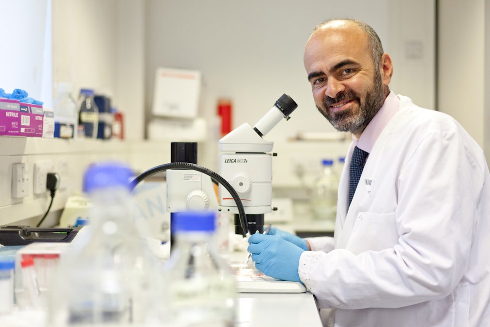
NIHR Professor Paolo de Coppi, senior author of the study from UCL Great Ormond Street Institute of Child Health and Great Ormond Street Hospital, said: “This is the first time that we’ve been able to make a functional assessment of a child’s congenital condition before birth, which is a huge step forward for prenatal medicine. Diagnosis is normally based on imaging such as ultrasound or MRI and genetic analyses.
“When we meet families with a prenatal diagnosis, we’re often unable to tell them much about the outcome because each case is different. We’re not claiming that we can do that just yet, but the ability to study functional prenatal organoids is the first step towards being able to offer a more detailed prognosis and, hopefully, provide more effective treatments in future.”
Researchers are able to monitor the microscopic organoids develop from fetal stem cells.
This UK arm of this work was carried out at the Zayed Centre for Research into Rare Disease in Children (ZCR). The ZCR, a partnership between GOSH, UCL and GOSH Charity brings together expertise in pioneering research and world-leading clinical care to drive new tests, treatments and cures for children with rare and complex diseases from lab bench to bedside.
This research was supported by the National Institute for Health and Care Research (NIHR) and Wellcome and all research at GOSH is underpinned by the NIHR GOSH Biomedical Research Centre.

